Portal Vein Embolization (PVE)
Portal vein embolization (PVE) is a procedure that induces regrowth on one side of the liver in advance of a planned hepatic resection on the other side. The procedure is frequently used in primary liver cancer (hepatocellular carcinoma) and colorectal liver metastases.
Harnessing the Unique Properties of the Liver
To be deemed a suitable surgical candidate, a patient must have enough functional liver remaining after the operation. The liver is a unique organ in that it can regrow after surgery, a property called "hypertrophy". However, the body requires that a minimum amount of liver remain (liver reserve) to support regrowth. Surgeons can safely remove up to 70% of the liver and expect full regeneration as long as the patient does not have substantial underlying liver disease such cirrhosis.
If the remaining liver reserve (liver remnant) is deemed insufficient to support liver regeneration, UCSF surgeons may use portal vein embolization to jump-start the regrowth of the liver remnant before the surgery.
How the Procedure Works
An interventional radiologist will place a needle percutaneously (through the skin) into the liver and identify the blood vessel on the side where the largest part of the tumor is being supplied. Tiny microspheres are then infused into the portal vein which supplies blood to the area, embolizing it by cutting off its blood supply.
This blockade of the blood supply induces the other side of the liver to regrow. After several weeks, the non-embolized side has grown enough so that surgery is now a viable option.
Novel Treatment of Bilobar Liver Cancer (HCC) Using PVE
Patients with tumors on both sides of the liver may require staged operations. For example, if the right side of liver has significantly more tumor volume than the left, hepatobiliary surgeons at UCSF will remove the entire right lobe.
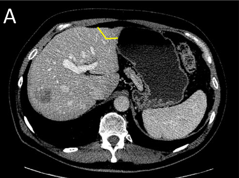
Surgeons first precondition the "future remnant" liver by resecting or ablating the tumors on the left side. This will become the functioning liver after the surgery. Surgeons then block the blood supply from the portal vein to the right liver for several weeks, promoting growth of the future left side remnant. The right liver is then completely removed or resected. This approach allows extra time for the left liver to regenerate, and reduces the risk of liver failure due to an insufficient amount of remnant liver.
 Case Study
Staged surgeries and portal vein embolization performed by hepatobiliary surgeons and interventional radiologists
 |
| (A) Staged Liver Resection: First Surgery (Stage I) - the tumors on the future remnant liver are treated first by resection or ablation |
 |
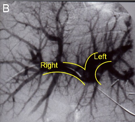
| (B) Before the next operation, a portal vein embolization is done: Venogram showing a normal portal vein prior to embolization |
 |
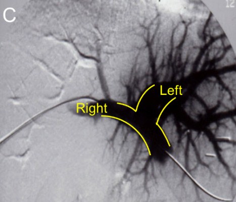
| (C) Loss of blood flow to the Right lobe of the liver after embolization |
 |
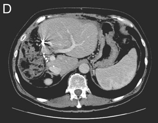
| (D) Stage II - Second Surgery: The right liver and tumors are removed. CT-Scan shows additional growth (hypertrophy) of the remaining liver remnant. |
